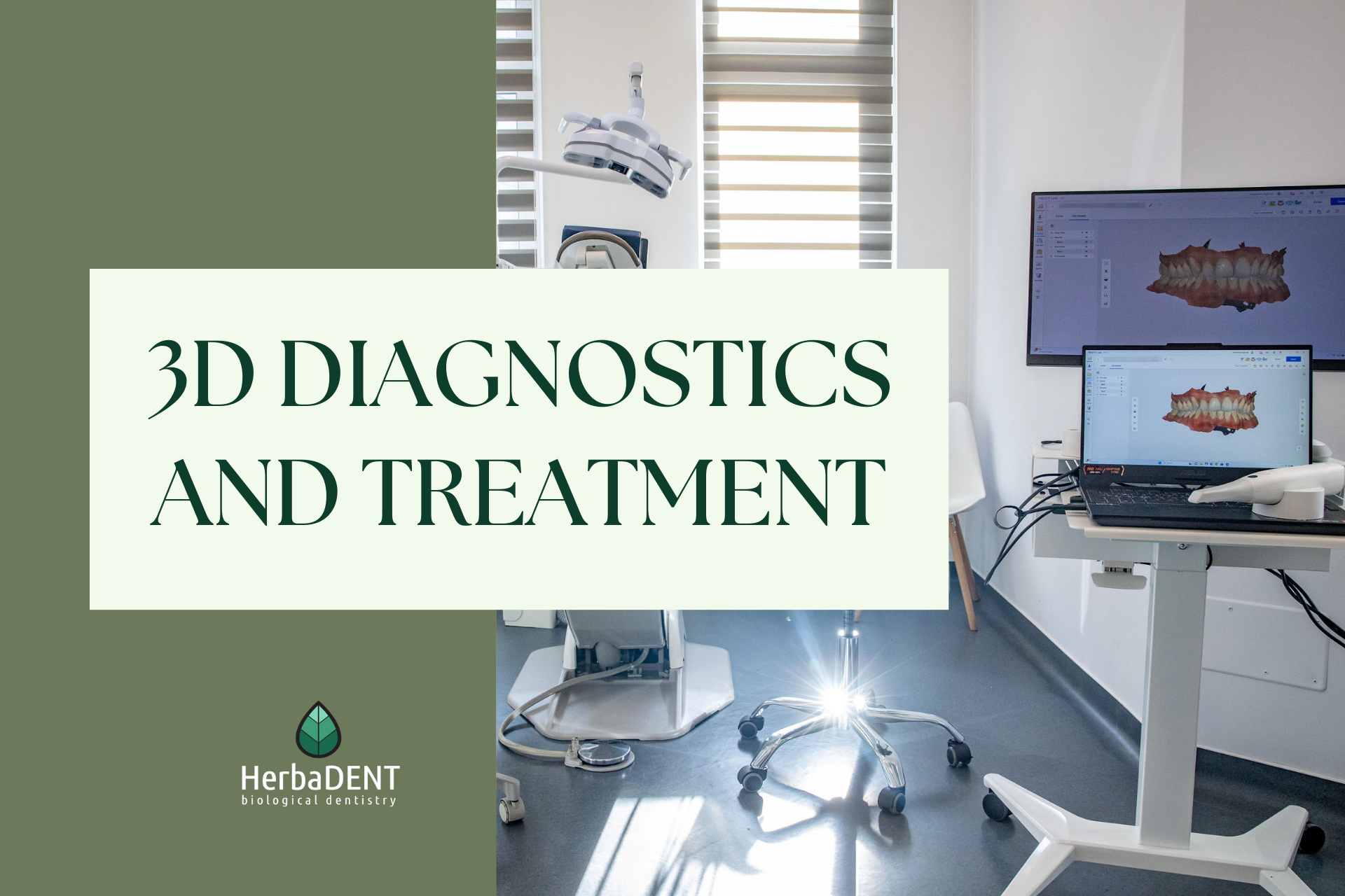
3D CBCT imaging diagnostic tests are often essential for establishing an accurate diagnosis and starting treatment.
The HerbaDENT Biological Dentistry clinic has one of the most modern dental CT equipment that allows accurate and reliable examinations, providing our colleagues with the highest level of medical care.
3D CBCT scans help the work of the dentist, with their help even latent, currently asymptomatic problems can be detected and more serious complications can be avoided.
What is dental CT?
Dental CT (computer tomograph) is practically an improved, modern version of the X-ray.
Imaging is carried out using X-rays in a transverse plane. In the resulting image there is no shadow or overlap, the area can be seen in 3D.
What is the difference between panoramic X-ray and CT?
A panoramic x-ray is a 2D image showing the denture, jaw bone and part of the facial cavity in an extended version. In the picture, the layers are projected on top of each other, “covered”.
In contrast, CT can also “see” areas that conventional X-rays do not. The resulting image is an accurate mapping of the patient. On the scans, the studied area can be orbited around, thanks to which even the smallest problems can be revealed.
In what cases can a dentist request a CT scan?
Thanks to the advanced technique, CT imaging may already be justified in almost all segments of dentistry. In some cases, after the scanning is completed, it is possible to establish an accurate diagnosis and determine the treatment, it can be an important starting point for preparing a treatment plan.
Root treatment:
With the help of the obtained images, it is possible to accurately see and determine the length, curvature and condition of the tooth root, as well as the extent of the existing inflammation.
We can also check previously treated teeth with the help of a CT scan to see at what stage the healing process is.
Primary cause investigation:
On CT scans, dental foci, cysts can be determined with great success.
Oral surgery:
It can be a key component of surgical interventions. With the help of imaging diagnostics, the tooth root and its position, the location of the tooth root in the bone, the running of nerves located in the jaw can be seen accurately.
Implantation:
Before installing implants, it is imperative to examine the existing bone and to assess its condition. With the help of scans, it is possible to accurately determine the type and size of the implant to be inserted. If the existing bone is insufficient, the necessary degree of bone supplement can also be determined on this basis.
Implantation can also be done using a digitally designed surgical template, the basis of which is also a CT scan. The computer program, analyzing the images, creates the surgical template, thanks to which the intervention can be much more accurate and faster, in addition, the recovery time can also be reduced. The finished template covers the area of intervention exactly to the millimetre, so the incision made during the operation can also be smaller.
Periodontology:
The degree of inflammation that has developed during gingivitis, periodontal disease and other diseases is clearly visible with the help of scans.
Dentures:
Before making dentures, it is important to examine the condition of the existing teeth, because the type of replacement that can be made for the absent teeth may depend on it. For example, in the case of a bridge, it is necessary to examine the existing pillar teeth to ensure that they can stably hold the finished denture.
How harmful is it to take a CT scan?
Modern equipment provides minimal X-ray radiation exposure to the body. One CT scan can replace several X-ray images.
Based on the above, it can be seen that dental CT scans form accurate images of the current condition of patients. With this, treatments can be precisely planned and executed. Imaging diagnostics is a help for both the dentist and the patient!

